Medial Collateral Ligament (MCL) Injury
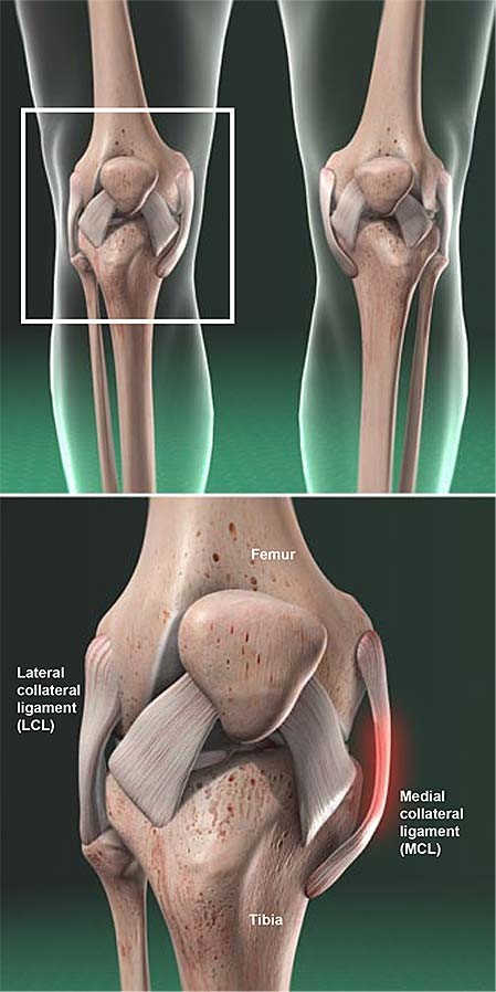
The knee joint allows the lower leg to flex or straighten. Four ligaments control and protect it.
- The medial collateral ligament (MCL) is located on the medial aspect of the knee or inside of the knee.
- The lateral collateral ligament (LCL) is located on the lateral aspect of the knee or outside of the knee.
- The anterior cruciate ligament (ACL) and posterior cruciate ligament (PCL) prevent the anterior and posterior movement of the knee joint.
Ligaments are tough bands of tissue that span a joint and attach to the bones on each side. The MCL is located on the inside of the knee and is attached to the femur and tibia. It holds the knee stable when a valgus stress is placed on the outer part of the leg.
The muscles surrounding the knee, especially the quadriceps muscles in the front of the thigh, and the hamstrings in the back of the thigh, also stabilize the knee joint.
Types of MCL Injuries
A sprain is an injury to a ligament. Healthcare providers grade knee ligament injuries by their severity.
- A grade I sprain refers to when one stretches but does not tear the fibers of the ligament.
- MCL tears are grade II sprains if the ligament fibers are partially torn.
- MCL tears are grade III sprains when the ligament is completely torn.
The MCL injury can also damage the medial meniscus and ACL.
Causes and Risk Factors of MCL Injuries
MCL injuries are the most common ligament sprains of the knee. They are common in sports and can occur in any age group.
Risks include contact sports and being male.
MCL injuries occur from a sudden impact on the outer part of the knee. The injury may be due to contact or noncontact activities.
Symptoms of an MCL Injury
Pain is the first symptom. It occurs along the course of the ligament and may be associated with swelling.
Diagnosis and Assessment of MCL Injuries
An MCL sprain is usually diagnosed through history and physical examination. The healthcare professional will ask about the injury and perform a physical examination, including touching the knee and assessing stability.
Plain X-rays and MRI may be used to visualize the MCL and assess for other injuries.
Treatment for an MCL Injury
MCL sprains heal with rest and physical therapy. Treatment may include wearing a knee sleeve or hinged knee brace, using crutches, and gradually returning to activity.
Surgery is considered in patients with additional injuries.
Home Remedies for MCL Injuries
Initial treatment includes rest, ice, compression, and elevation. Using crutches and over-the-counter anti-inflammatory medications may help with pain control.
Prognosis and Complications of MCL Injuries
Athletes can usually return to play within a few weeks. Grade III sprains may require a longer recovery period.
The major complication is instability, which may require surgery after initial therapy.
Prevention of MCL Injuries
MCL sprains cannot be completely prevented, but avoiding traumatic contact can reduce the risk. The use of braces for protection is controversial.
By clicking Submit, I agree to the MedicineNet’s Terms & Conditions & Privacy Policy and understand that I may opt out of MedicineNet’s subscriptions at any time.
Donatelli, R.A., and M.J. Wooden. Orthopedic Physical Therapy, 4th Ed. St. Louis, Mo.: Churchill Livingstone, 2009
Miller, Seamon J. "Medial Collateral Ligament Injuries of the Knee." MRI-Arthroscopy Correlations. Ed. S. Brockmeier. New York: Springer, 2015.
Roach, C.J., C.A. Haley, K.L. Cameron, M. Pallis, S.J. Svoboda, and B.D. Owens. "The Epidemiology of Medial Collateral Ligament Sprains in Young Athletes." Am J Sports Med 42.5 May 2014: 1103-1109.


