Contents
- 1 Iritis
- 1.0.1 What is the difference between uveitis vs. iritis?
- 1.0.2 What causes iritis?
- 1.0.3 What are signs and symptoms of iritis?
- 1.0.4 How do healthcare professionals diagnose iritis?
- 1.0.5 What are the treatment options for iritis?
- 1.0.6 What is the prognosis for iritis?
- 1.0.7 How long does iritis last?
- 1.0.8 What are complications of iritis?
- 1.0.9 What is the latest research on iritis?
- 1.0.10 Where can I find more information about iritis?
Iritis
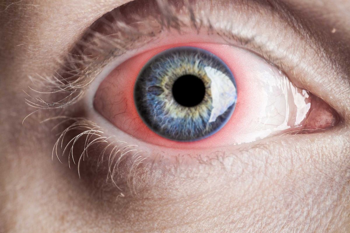
Iritis is inflammation of the iris, the colored portion of the eye around the pupil. It causes redness, pain, sensitivity to light, tearing, and blurred vision. It usually affects one eye, but can affect both in some systemic diseases.
In rare cases, iritis can lead to serious eye damage and permanent vision loss.
An ophthalmologist must investigate the causes of iritis and promptly initiate appropriate treatment. Healthcare providers can easily treat iritis without causing any damage.
What is the difference between uveitis vs. iritis?
The uvea is a tissue in the eye that consists of three parts: the iris, the ciliary body behind the iris, and the choroid at the back of the eye.
- Uveitis refers to swelling along the uveal tract.
- Iritis is another name for anterior uveitis or iridocyclitis.
What causes iritis?
Iritis can have many causes. In some cases, the inflammation is idiopathic (of unknown cause), and the acute iritis may occur only once in a person’s life.
Iritis can be associated with various conditions, including systemic diseases. In those cases, it often recurs.
- Infections such as herpes simplex virus, herpes zoster virus (shingles), tuberculosis, syphilis, etc.
- Eye injury resulting in traumatic iritis. In rare cases, previous eye trauma can cause delayed onset iritis in the non-traumatized eye.
- Autoimmune disorders like juvenile rheumatoid arthritis, HLA-B27-associated diseases (e.g. ankylosing spondylitis), and collagen vascular diseases (e.g. lupus)
- Inflammation after eye surgery
- Sarcoidosis
- Adamantiades-Behçet’s disease, which causes inflammation around the blood vessels in the uveal tissue
- Inflammatory bowel disease, specifically ulcerative colitis and Crohn’s disease
- Certain medications, including prostaglandin analog glaucoma medications
- More posterior uveitis (e.g. intermediate uveitis and choroiditis) with inflammatory cells spilling over into the anterior chamber (the front part of the eye), which can mimic iritis. Similarly, retinal detachment can cause pigment and cell spillover into the anterior chamber, mimicking iritis as well.
QUESTION
What are signs and symptoms of iritis?
Symptoms of iritis may include:
- Eye pain
- Blurred vision, especially with extensive posterior uveitis
- Sensitivity to light and pain when exposed to bright light (photophobia)
Signs of iritis may include:
- Redness
- Tearing
- Presence of inflammatory white blood cells in the anterior chamber, visible under the slit lamp microscope
- A hypopyon may form, which is a collection of white blood cells settling in the bottom half of the anterior chamber. Sometimes the hypopyon is visible with the naked eye as a layer of white material in front of the inferior iris. Traumatic iritis can also cause a collection of red blood cells called a hyphema in a similarly located area.
- Possible change in eye pressure
How do healthcare professionals diagnose iritis?
An eye doctor will obtain a patient’s complete medical and ocular history, including any family history of uveitis. They will perform a thorough eye exam to look for iritis, complications, and clues to determine the cause.
The physician will use a slit lamp microscope to confirm the diagnosis, looking for white cells in the aqueous (the eye’s liquid) to indicate inflammation. A complete eye examination, including dilation to examine the back of the eye, helps determine the extent of the inflammation (iritis vs. more extensive uveitis) and provides clues to the cause.
If no obvious cause is found, and it is the first occurrence, additional testing may not be necessary. However, if the iritis is severe or recurring or if posterior uveitis is present, the doctor will order additional tests, such as blood work and a chest X-ray, to rule out associated diseases.
If the eye pressure is dangerously high, it needs to be addressed along with the iritis.
What are the treatment options for iritis?
Steroid eye drops are the main treatment. In severe cases, steroids may need to be injected into the eye or taken orally.
- If an autoimmune disease is associated with iritis, drugs like immunomodulatory therapy (e.g. methotrexate, azathioprine, mycophenolate) and biologic response modifiers (e.g. infliximab, adalimumab) used to treat the disease may help resolve the iritis.
- If an infection causes the iritis, anti-infectives (antibiotics, antivirals, antifungals, antiparasitics, etc.) are necessary.
An eye doctor may also prescribe a cycloplegic (dilating drop) like cyclopentolate to relieve pain. Dilation can prevent the swollen iris from adhering to the lens behind the pupil.
If portions of the iris become stuck to the lens, synechiae can form. Too many synechial attachments can cause dangerously high eye pressure and glaucomatous vision loss. To reduce eye pressure (due to iritis or synechiae), pressure-lowering eyedrops are prescribed.
What is the prognosis for iritis?
In most cases, iritis responds well to a short course of steroid eye drops and cycloplegics (dilation drops), resulting in no complications. However, the prognosis ultimately depends on the severity, frequency, and duration of the iritis and any complications.
Associated diseases or infections can cause eye damage leading to chronic pain or vision loss.
How long does iritis last?
Usually, iritis clears in days but may last for months or become chronic and recurrent. Immediate recognition and treatment by a physician are crucial.
Patients should continue treatment until the inflammation completely resolves to avoid complications associated with chronic iritis or uveitis.
What are complications of iritis?
Some complications of iritis may include:
- Permanent vision loss: Rare but can occur if the retina develops fluid collections known as CME (cystoid macular edema) or if high eye pressure leads to glaucomatous damage to the optic nerve.
- Scarring of the iris: Synechiae formations can cause acutely or chronically elevated eye pressures, leading to glaucoma. Scarring can also be a side effect of the steroids used to treat iritis. Steroid use can also contribute to premature cataract formation.
- Patients with longstanding iritis may develop corneal mineral deposits (band keratopathy), resulting in blurred vision and dry eye symptoms. Chronically elevated eye pressure can damage the corneal endothelial cells at the back of the cornea, causing corneal cloudiness.
What is the latest research on iritis?
- Major research is ongoing in the field of uveitis, which includes various inflammatory eye conditions, including iritis.
- Research primarily focuses on managing severe cases or cases involving more extensive eye involvement, as iritis generally responds well to treatment. This research improves our understanding of the mechanism of iritis and its treatment.
- Research is also being conducted to find more effective medications and the best way of delivering them to the eye.
Where can I find more information about iritis?
The American Academy of Ophthalmology: http://www.aao.org
NIH: National Eye Institute: http://www.nei.nih.gov/
By clicking Submit, I agree to the MedicineNet’s Terms & Conditions & Privacy Policy and understand that I may opt out of MedicineNet’s subscriptions at any time.
Foster, C.S., et al. "The Ocular Immunology and Uveitis Foundation preferred practice patterns of uveitis management." Surv Ophthalmol 61.1 Jan.-Feb. 2016: 1-17.
Foster, C.S., et al. "The Ocular Immunology and Uveitis Foundation preferred practice patterns of uveitis management." Surv Ophthalmol 61.1 Jan.-Feb. 2016: 1-17.


