Contents
CT Scan (Computerized Tomography, CAT Scan)
Computerized tomography (CT), formerly known as computerized axial tomography (CAT) scan, combines X-ray images with a computer to generate cross-sectional and three-dimensional views of the body’s internal organs and structures. It is commonly referred to as a CT scan. This procedure is used to identify normal and abnormal structures in the body, assist in procedures, and guide the placement of instruments or treatments.
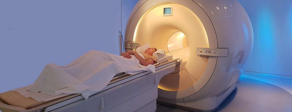
A donut-shaped X-ray machine takes images of the body from different angles. These images are processed by a computer to create cross-sectional pictures. Each picture represents a "slice" of the body, similar to slicing a loaf of bread. When these slices are combined, a three-dimensional picture of an organ or abnormal structure can be obtained.
Purpose of a CT Scan
CT scans analyze internal structures in various parts of the body. They can identify traumatic injuries, tumors, infections, and evaluate bone density. Contrast material may be used to enhance scans and visualize specific structures. CT scans can also verify the presence of tumors, infection, abnormal anatomy, or changes caused by trauma in the abdomen.
This technique is painless and provides accurate images that guide radiologists in performing procedures such as biopsies, fluid removal, and abscess drainage.
Risks of a CT Scan
A CT scan is a low-risk procedure. Adverse reactions to contrast material are rare and usually self-limiting. In rare cases, severe allergic reactions can occur, but they can be treated. Kidney toxicity from the contrast material is extremely rare with newer agents.
Radiation exposure during a CT scan is minimal and has no proven adverse effects in men and nonpregnant women. Pregnant women should discuss alternative imaging methods with their doctor to avoid potential risks to the fetus. Lifetime cancer risk from CT scan radiation is small but more significant for children.
Preparing for a CT Scan
Prior to a CT scan, patients may need to avoid food and fluids, especially when contrast material is used. Metallic materials and certain clothing should be removed to ensure clear images. Patients are placed on a movable table and stay still during the scan. CT scan technology is continually improving, offering faster and more accurate visualization of internal organs.
References:
1. Braunwald, Eugene, et al. Harrison’s Principles of Internal Medicine, 15th Ed. McGraw-Hill, 2010.
2. FDA.gov. Computed Tomography.
3. Tramma, Simone, et al. "Helical CT Scans and Lung Cancer Screening." CDC NIOSH Science Blog. 10 Jan. 2011.


