Contents
Bunions (Hallux Valgus)
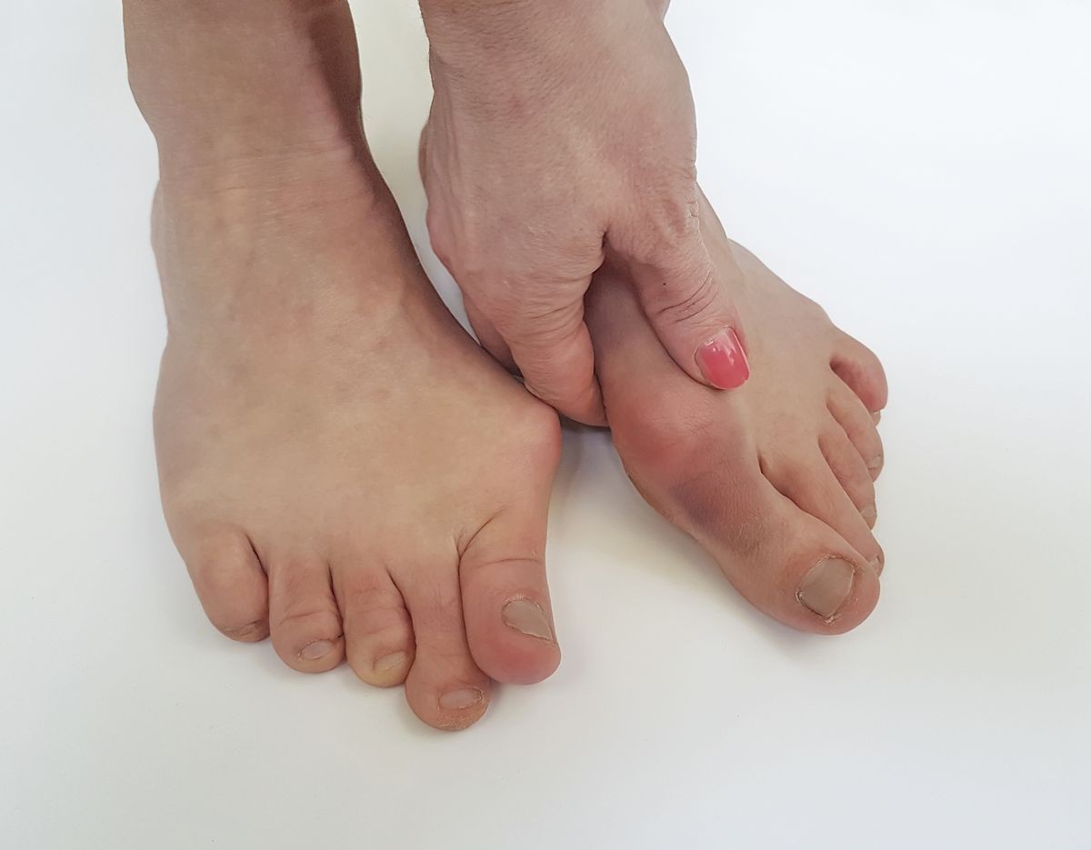
The common bunion is a localized area of enlargement or prominence of the inner portion of the joint at the base of the big toe. The enlargement represents a misalignment of the big toe joint (metatarsal phalangeal joint) and, in some cases, additional bone formation. The misalignment causes the big toe to point outward and rotate (medically termed hallux abducto valgus deformity) toward the smaller toes. This deformity is progressive and will increase with time although the symptoms may or may not be present.
The enlarged joint at the base of the big toe (the first metatarsophalangeal joint, or MTP joint) can become inflamed with redness, tenderness, and pain. A small fluid-filled sac (bursa) adjacent to the joint can also become inflamed (bursitis), leading to additional swelling, redness, and pain. Deeper joint pain may occur as localized arthritis develops in later stages of the deformity.
A less common bunion is located at the joint at the base of the fifth (pinky) toe. This bunion is sometimes referred to as a tailor’s bunion or bunionette. The misalignment at the fifth MTP joint causes the pinky toe to point inward and create an enlargement of the joint.
What causes bunions?
While the precise cause is not known, bunions are believed to be caused by multiple factors including abnormal foot function and mechanics, such as overpronation, abnormal anatomy at first MTP joint, and genetic factors. Overpronation is a common cause of bunion in younger individuals. Abnormal biomechanics can lead to instability of the metatarsal phalangeal joint and muscle imbalance, resulting in the deformity. Abnormal anatomy at the first MTP joint can predispose an individual to develop a bunion deformity. A study has found a significant genetic heritability of bunion deformity among people of European descent.
Although shoe gear doesn’t directly cause a bunion, it can certainly make the condition painful and swollen. Other less common causes of bunion deformities include trauma, neuromuscular disorders, and limb-length discrepancies.
Various arthritic conditions can cause or exacerbate bunion deformity. Gout and rheumatoid arthritis often cause severe bunions to develop along with other toe deformities.
Bunions most commonly affect women. Some studies report that bunion symptoms occur nearly 10 times more frequently in women. It has been suggested that tight-fitting shoes, especially high-heel and narrow-toed shoes, might increase the risk for bunion formation. Tight footwear certainly is a factor in precipitating the pain and swelling of bunions.
Other risk factors for the development of bunions include abnormal formation of the bones of the foot at birth and arthritic diseases such as rheumatoid arthritis. In some cases, repetitive stresses to the foot can lead to bunion formation. Bunions are common in ballet dancers.
IMAGES
What are the symptoms of bunions?
Bunions may or may not cause symptoms. Early symptoms may be foot pain in the involved area when walking or wearing shoes; rest and/or change to a wider shoe relieves this pain. Shoe pressure in this area can cause intermittent pain while the development of arthritis in more severe bunions can lead to chronic, constant pain. For arthritis sufferers, removing shoes or resting does not always provide relief. Besides ill-fitting shoes, unsupportive flimsy soled shoes can add stress to the bunion joint and increase pain and instability to the area.
Skin changes such as blisters and calluses may also develop as a result of repeated friction and pressure on the prominence by ill-fitting shoes or from the deformity caused by arthritis.
Bunions that cause marked pain are often associated with swelling of the soft tissues, redness, joint stiffness, and local tenderness. It is important to note that, in post-pubertal men and postmenopausal women, pain at the base of the big toe can be caused by gout and gouty arthritis that is similar to the pain caused by bunions.
Diagnosis of bunions
A physician will consider a bunion as a possible diagnosis when noting the symptoms described above. The anatomy of the foot, including joint and foot function, is assessed during the examination. Radiographs (X-ray films) of the foot can be helpful to determine the integrity of the joints of the foot and to screen for underlying conditions, such as arthritis or gout. X-ray films are an excellent method of calculating the alignment of the toes when taken in a standing position (weight-bearing).
What is the treatment for bunions?
Nonsurgical treatments such as rest and wearing loose (wider) shoes or sandals with a supportive sole can often relieve the irritating pain of bunions. Walking shoes may have advantages over high-heeled styles that pressure the sides of the foot.
Anti-inflammatory medications can help ease inflammation and provide pain relief. Local cold-pack application is sometimes helpful, as well.
Bunion shields or pads can reduce pressure on the bunion caused by closed shoes. A bunion splint may also help if the condition is still in its early stage with minimal deformity. Splints help to place the toe in a corrected position, while stretching tight soft tissue structures. Night splints are used while sleeping and splint booties can be worn inside a shoe. Taping and toe separators can also be used to achieve the same goal.
Stretches and manipulation of first MTP joint structures can help preserve joint mobility. This can delay onset of arthritis at the joint.
Custom orthotics are used to improve foot mechanics and address overpronation. Orthotics can prevent progression of bunion and relieve pain by improving joint function. In addition, orthotics need to be worn in stable, well-fitting, laced shoes.
Inflammation of the joint at the base of the big toe can often be relieved by a local injection of cortisone.
Constant pressure or friction can lead to skin breakdown and infection that may require antibiotic therapy.
When the measures above are effective in relieving symptoms, patients should avoid irritating the bunion again by optimizing footwear and foot care.
Is surgery needed for bunions?
A surgical operation is considered for correction of the bunion if conservative care does not provide sufficient pain relief. The surgical operation to cure a bunion is referred to as a bunionectomy. Surgical procedures can correct the deformity and relieve pain, leading to improved foot function. These procedures typically involve removing bony growth of the bunion while realigning the big toe joint. Surgery is often, but not always, successful.
What is the prognosis for bunions?
The treatments described above are very effective in treating bunion deformities, and the prognosis can be excellent. However, the correct diagnosis is essential to define any underlying associated deformities as well as the bunion severity. Also, a bunion is a progressive deformity and will get worse with time. It can cause instability to the rest of the foot and sometimes lead to arthritis in the joint at the base of the big toe. It is advised to consult with a foot specialist to fully evaluate a bunion. Early recognition, diagnosis, and bunion care can prevent debilitating aftereffects.
Is it possible to prevent bunions?
If the diagnosis is made early on, such as in preadolescence, bunion development can be slowed and, in some cases, arrested with the proper supportive shoe gear and custom functional shoe inserts. Physical therapy involving stretch can also be beneficial. Avoidance of certain athletic activities with improper shoe fit and toe pressure can prevent the symptoms that occur with bunions. Early examination by a podiatrist is recommended.
"Adult Hallux Valgus." Surgery of the Foot and Ankle. Eds. Mann, R., and M. Coughlin. St. Louis: Mosby, 1999: 150.
Brantingham, J.W., S. Guiry, H.H. Kretzmann, et al. "A pilot study of the efficacy of conservative chiropractic protocol using graded mobilization, manipulation and ice in the treatment of symptomatic hallux abductovalgus bunion." Clin Chiro 8 (2005): 117.
du Plessis, M., B. Zipfel, J.W. Brantingham, et al. "Manual and manipulative therapy compared to night splint for symptomatic hallux abducto valgus: an exploratory randomised clinical trial." Foot (Edinb) 21 (2011): 71.
Ferrari, J., and J. Malone-Lee. "The shape of the metatarsal head as a cause of hallux abductovalgus." Foot Ankle Int 23 (2002): 236.
"Forefoot deformity caused by abnormal subtalar joint pronation." Normal and Abnormal Functions of the Foot, Clinical Biomechanics, Vol. 2. Eds. Root, M.L., W.P. Orien, and J.H. Weed. Los Angeles: Clinical Biomechanics Corporation, 1977: 376.
Fritz, G.R., and D. Prieskorn. "First metatarsocuneiform motion: a radiographic and statistical analysis." Foot Ankle Int 16 (1995): 117.
Haas, C., B. Kladny, S. Lott, et al. "Progression of foot deformities in rheumatoid arthritis–a radiologic follow-up study over 5 years." Z Rheumatol 58 (1999): 351.
Hannan, M. T., H.B. Menz, J.M. Jordan, L.A. Cupples, C.H. Cheng, and Y.H. Hsu. "High Heritability of Hallux Valgus and Lesser Toe Deformities in Adult Men and Women." Arthritis Care & Research 65 (2013): 1515-1521.
Klippel, John H., eds., et al. Primer on the Rheumatic Diseases, 13th Ed. New York: Springer, 2008.
Phillips, D. Biomechanics in Hallux Valgus and Forefoot Surgery. New York: Churchill Livingstone, 1988: 39.
Tanaka, Y., Y. Takakura, T. Takaoka , et al. "Radiographic analysis of hallux valgus in women on weightbearing and nonweightbearing." Clin Orthop Relat Res 1997: 186.
Torkki, M., A. Malmivaara, S. Seitsalo, et al. "Surgery vs orthosis vs watchful waiting for hallux valgus: a randomized controlled trial." JAMA 285 (2001): 2474.


