Contents
- 1 When Do You Need a Target Scan While Pregnant?
- 1.0.1 3 main objectives of a target scan
- 1.0.2 12 most commonly detected abnormalities in a target scan
- 1.0.3 Reasons for missing identification of abnormalities during a target scan
- 1.0.4 How is the target scan done?
- 1.0.5 How to prepare for a target scan
- 1.0.6 What is the importance of a target scan?
- 1.0.7 Things to know about a target scan
When Do You Need a Target Scan While Pregnant?
A target scan is a crucial prenatal ultrasound that assesses fetal development, location, and detects impairments.
A target scan is also known as targeted imaging for fetal anomalies scan or fetal anomaly scan. This scan is required during the second trimester of pregnancy, between 18 and 20 weeks of gestation. An early target scan can be done at 16 weeks of pregnancy.
This scan monitors the baby’s physical development, growth, and detects any anatomical abnormalities.
3 main objectives of a target scan
- To predict the baby’s structural integrity
- To detect serious and potentially fatal anomalies
- To suggest the possibility of an abnormality, requiring additional testing
Target scan examines your child’s natural and healthy growth. While it can identify significant abnormalities, it cannot detect all defects. Even if your baby appears healthy in the scan, there is still a possibility of birth defects.
12 most commonly detected abnormalities in a target scan
The twelve most commonly detected abnormalities in a target scan are:
- Anencephaly (98 percent): Abnormal brain and skull bone growth, resulting in newborn fatality. Incidence: 1 in 2,000 babies
- Gastroschisis (98 percent): Defect in the abdominal wall, causing intestine growth outside the baby’s abdomen. Newborn fatality. Incidence: 1 in 2,000 babies
- Patau’s syndrome (Trisomy 13) (95 percent): Rare chromosomal disease with three copies of chromosome 13. Incidence: 1 in 4,000 babies
- Edwards’ syndrome (Trisomy 18) (95 percent): Rare chromosomal disease with three copies of chromosome 18. Incidence: 1 in 1,500 babies
- Open spina bifida (90 percent): Gap or split in the spine, affecting spinal cord growth. Incidence: 1 in 1,666 babies
- Bilateral renal agenesis (84 percent): Kidneys have not formed, resulting in newborn fatality. Incidence: 1 in 5,000 babies
- Exomphalos (80 percent): Failure of abdomen to close, causing organ emergence. Incidence: 1 in 2,500 babies
- Cleft lip (75 percent): Incomplete connection of baby’s lips. Incidence: 1 in 1,300 babies
- Lethal skeletal dysplasia (60 percent): Bone and cartilage disorders affecting fetal skeleton development. Incidence: 1 in 10,000 babies
- Congenital diaphragmatic hernia (60 percent): Underdeveloped diaphragm, separating organs. Incidence: 1 in 2,500 babies
- Congenital heart diseases (50 percent): Various cardiac defects that may require medical treatment. Incidence: 1 in 125 with critical defects in 1 in 500 babies
- Down syndrome (Trisomy 21) (50 percent): Genetic disorder with additional chromosome 21. Incidence: 1 in 700 babies
QUESTION
Reasons for missing identification of abnormalities during a target scan
Common reasons for missing the identification of some anomalies during an ultrasound scan are:
- Unideal fetal position for identifying body parts
- Mother overweight
- Presence of obstructive fibroids
In such cases, a scan at 23 weeks is done. If the scan cannot be completed, your baby will receive a full examination following birth.
How is the target scan done?
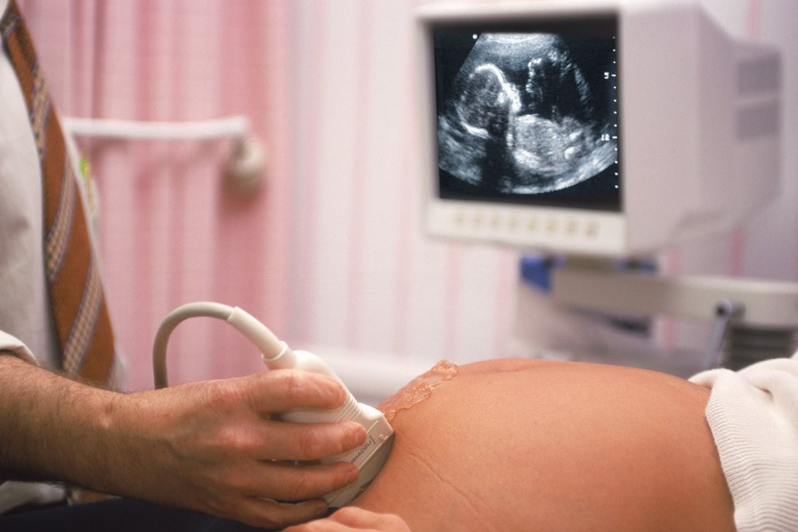
The scanning procedure is swift and painless. The sonographer or radiologist applies gel to your belly and moves a handheld probe over the area. The gel ensures good contact with your skin.
You will see images of your baby on the ultrasound screen. The sonographer may apply slight pressure to your abdomen to obtain clear photos, which may be uncomfortable but safe.
- The complete procedure lasts 30 to 40 minutes. Delays may occur if the baby moves or the sonologist cannot check an area due to the baby’s posture.
- The sonologist may ask you to wait for the baby’s right position or call you back another day to continue the inspection.
- Drinking water before the scan may improve visibility, though it is not always necessary.
How to prepare for a target scan
Before a target scan, remember the following:
- Bring your obstetrician’s sonography prescription.
- Bring your prior ultrasound scan records if available.
- A full bladder is not required.
- Allow enough time as this scan may take longer.
What is the importance of a target scan?
A target scan is one of the most important prenatal ultrasound scans.
- The scan assesses fetal development, location, and detects impairments.
- This scan aids in the detection of anomalies and helps you make decisions about the pregnancy in extreme cases.
This scan is a personal choice but highly recommended by prenatal experts.
The target scan is done between 18 and 20 weeks of pregnancy when the baby grows around six inches. It captures abnormalities during anatomical development.
While many abnormalities may go undetected, approximately 50 percent of Down syndrome problems and congenital heart defects can be identified.
- The target scan evaluates fetal development, health, and anatomy.
- The scan determines fetal size, weight, and location of placenta, umbilical cord, and amniotic fluid.
- Improper placenta location can lead to complications.
In some cases, fetal organs may not develop normally, which can lead to fetal demise during pregnancy or shortly after birth. Early detection allows your obstetrician to provide appropriate steps, medications, and reassurance for the rest of the pregnancy.
Things to know about a target scan
- Scientific studies confirm the safety of ultrasound imaging when conducted by experts.
- The sonographer may identify the baby’s sex, but it may be incorrect in some cases.


