Contents
- 1 Four Main Functions of the Heart
- 1.0.1 Medical Conditions Related to the Heart
- 1.0.2 Difference Between Left and Right Heart Catheterization
- 1.0.3 Why is Cardiac Catheterization Done?
- 1.0.4 Who Should Not Undergo Cardiac Catheterization?
- 1.0.5 Preparing for Cardiac Catheterization
- 1.0.6 What Happens During Cardiac Catheterization?
- 1.0.7 What Happens After Cardiac Catheterization?
- 1.0.8 How Serious is Cardiac Catheterization?
Four Main Functions of the Heart
The heart is a muscular organ located in the chest, just behind and slightly to the left of the breastbone. It is roughly the size of a closed fist. The heart continuously pumps blood through a network of blood vessels called arteries and veins. The heart and its blood vessels are part of the cardiovascular system.
The heart has four chambers: the upper chambers are called atria, and the lower chambers are known as ventricles. The right atrium and right ventricle are referred to as the right heart, while the left atrium and left ventricle are referred to as the left heart. These chambers are separated by partitions, called septum.
- The right atrium receives deoxygenated blood from the body and pumps it to the right ventricle.
- The right ventricle receives blood from the right atrium and pumps it to the lungs to load it with oxygen.
- The left atrium receives oxygenated blood from the lungs and pumps it to the left ventricle.
- The left ventricle, the strongest chamber of the heart, pumps oxygen-rich blood to the rest of the body.
The flow of blood into, within, and from the heart is controlled by four valves. The heart itself is supplied with nutrients and oxygen through coronary arteries. Nerve tissue facilitates the heart’s rhythmic heartbeat. The heart is protected by a fluid-filled sac called the pericardium, which prevents friction between the heart and surrounding organs.
Medical Conditions Related to the Heart
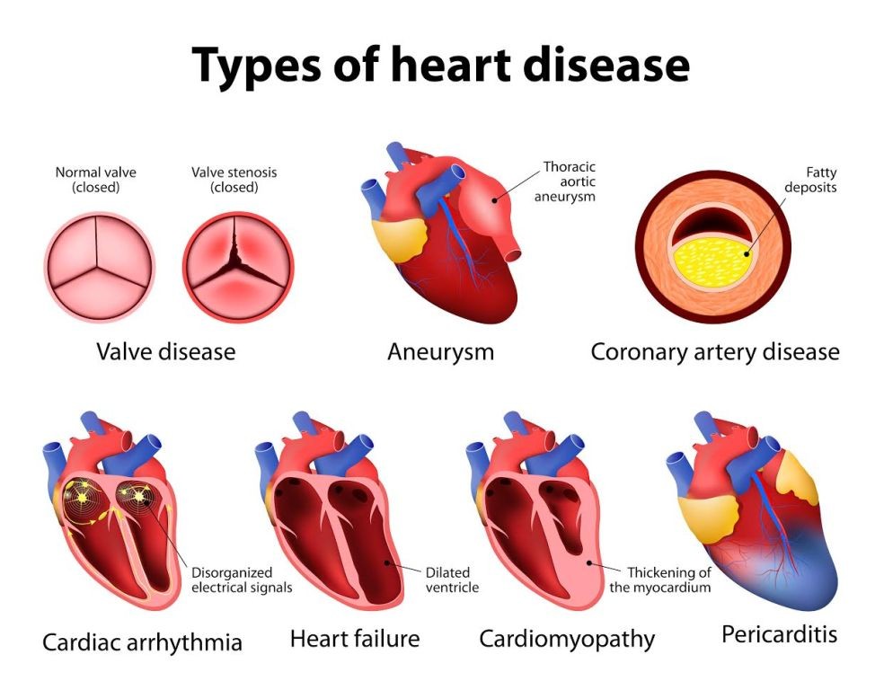
Some common heart conditions include:
- Coronary artery disease (CAD): Narrowing of the arteries supplying blood to the heart; complete blockage can lead to a heart attack.
- Stable angina pectoris: Chest pain due to insufficient blood supply to the heart during physical activity; relieved by rest.
- Unstable angina pectoris: New or worsening chest pain, even at rest; requires urgent medical attention.
- Myocardial infarction (heart attack): Death of heart muscle due to a sudden blockage in a coronary artery.
- Arrhythmia (dysrhythmia): Abnormal heart rhythm that affects the heart’s ability to pump blood effectively.
- Congestive heart failure (CHF): Inability of the heart to pump blood to the body efficiently, leading to fluid collection.
- Cardiomyopathy: Disease that enlarges, thickens, and weakens the heart, impairing its ability to pump blood.
- Myocarditis: Inflammation of the heart muscle.
- Pericarditis: Inflammation of the membrane surrounding the heart (pericardium).
- Pericardial effusion: Collection of fluid between the pericardium and the heart itself.
- Heart valve diseases: Disorders that affect the valves directing blood flow into and out of the heart.
- Cardiac arrest: Sudden stoppage of heart function.
IMAGES
Difference Between Left and Right Heart Catheterization
Left heart catheterization goes through the artery, while right heart catheterization goes through the veins.
Cardiac catheterization, also known as cardiac cath or heart cath, is a procedure used to examine heart function.
A catheter, a thin tube, is inserted into a blood vessel in the arm or leg and guided to the heart’s arteries using an X-ray camera. Contrast dye is then injected into the blood vessel through the catheter to obtain X-ray images of the valves, arteries, and heart chambers.
- Left heart catheterization involves passing the catheter through an artery.
- Right heart catheterization involves passing the catheter through a vein.
Why is Cardiac Catheterization Done?
Cardiac catheterization is performed to diagnose heart conditions such as:
- Atherosclerosis: Deposits of fat, cholesterol, calcium, and fibrin (clotting materials) that clog arteries.
- Cardiomyopathy: Enlargement of the heart due to muscle thickening or weakening.
- Congenital heart disease: Structural defects in the heart present since birth.
- Heart failure: Weakened heart muscles resulting in poor blood pumping and congestion in blood vessels and lungs.
- Heart valve disease: Failure of one or more heart valves leading to reduced blood flow within the heart.
- Evaluation of coronary artery disease (CAD) in patients with unclear clinical symptoms.
Who Should Not Undergo Cardiac Catheterization?
Patients with the following conditions should not undergo cardiac catheterization:
- Severe uncontrolled blood pressure
- Severe anemia
- Kidney failure
- Allergy to contrast dyes
- Stroke
- Severe gastrointestinal bleeding
- Electrolyte abnormalities
- Impaired blood clotting
- Ventricular arrhythmia
- Untreated infection or unexplained fever
Preparing for Cardiac Catheterization
Your physician will explain the procedure, including risks and benefits. You will also be instructed to:
- Sign an informed consent
- Follow specific dietary restrictions 24 hours before the test
- Fast for six to eight hours prior to the procedure
- Inform the doctor about any allergies to dyes used in the procedure
- Provide medical and medication history, especially any drug allergies
- Discontinue certain medications before the procedure
- Arrange for someone to accompany you during the procedure
What Happens During Cardiac Catheterization?
- Prior to the procedure, a nurse will administer sedatives through an intravenous (IV) line to help you relax.
- Your doctor will use local anesthetic to numb the area where the catheter will be inserted.
- The groin area will be cleaned and shaved. The doctor will use a needle to access the blood vessel.
- An introducer sheath will be inserted through which the catheter will be advanced to the blood vessels. You may feel pressure, but notify the doctor of any pain.
- A small amount of dye will be injected into the arteries once the catheter reaches them.
- An X-ray camera will capture images of your arteries and heart chambers. The catheter will be gradually removed after the procedure.
What Happens After Cardiac Catheterization?
You will be taken to the recovery room for a few hours. During this time:
- Pressure will be applied to the puncture site to stop bleeding.
- You will need to lie flat on the bed.
- Your vital signs, such as blood pressure and pulse, will be monitored during recovery.
- Inform the doctor of any swelling, pain, or bleeding at the puncture site.
How Serious is Cardiac Catheterization?
Cardiac catheterization is generally considered safe, but like any procedure, it carries risks, including:
- Heart attack
- Blood clots
- Infection
- Kidney damage
- Stroke
- Bruises
- Bleeding
- Arrhythmia
- Allergic reaction to contrast dye
- Air embolism
By clicking "Submit," I agree to the MedicineNet Terms and Conditions and Privacy Policy. I also understand that I may opt out of MedicineNet subscriptions at any time.


